A PFO can be closed without the need for surgery, using a small umbrella-like device. The closure device is passed to the heart using a small tube (catheter) inserted in a vein at the top of the thigh.
This closure method is very safe and effective. The procedure can be done with mild sedation and local anesthesia and usually takes about 30 minutes to complete. Patients typically go home the next day and are back to all their usual activities in a few days.
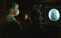
PFO closure is done in the cardiac catheterization laboratory where special x-ray and ultrasound equipment are used to guide placement of the closure device.
Mount Sinai has recently undertaken a major renovation of the cardiac catheterization suite, installing the most-advanced imaging equipment available.
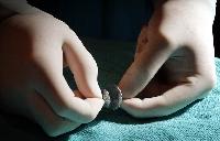
PFO closure device
An Amplatzer PFO Occluder
An occluder used to close PFOs is a small umbrella-like device that can be compressed inside a small tube (catheter), placed at the desired location in the heart, and then opened.
The size of the occluder chosen is based on the individual patient anatomy.
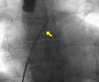
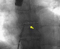
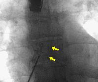
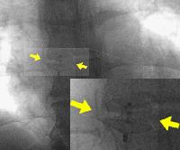
The following illustrates the method of PFO closure. These are actual x-ray images taken during a PFO closure. The position of the closure device and catheter is monitored by special x-ray and ultrasound equipment.
The delivery catheter is threaded from the vein at the top of the thigh up to the heart.
The catheter is then placed through the PFO.
The PFO occluder is collapsed down into the delivery catheter.
The left atrial disk is then opened in the left atrium and then pulled back to the flap of the PFO.
The right atrial disk is then opened. The PFO is now closed. The device position is verified by special ultrasound equipment to ensure that the device is in the correct position. Note that the device is still attached to the delivery cable and can be removed or repositioned if the physician is unsatisfied with the device position.
When the physician is satisfied with the device position, the occluder is released from the delivery cable and the occluder assumes its final position in the PFO. A final check is made by ultrasound, and the procedure is complete. The entire procedure takes about 30 minutes.
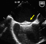
This is the echocardiogram image (special ultrasound) showing the Amplatzer PFO Occluder in-position closing the PFO.
What What to expect on the day of the procedure
The patient arrives to the catheterization laboratory and is checked in. An IV is placed and any needed bloodwork is drawn. The patient is then taken to the catheterizaiton suite. The femoral area is cleaned and draped are placed to keep the area clean. Intravenous sedation is administered to lessen any anxiety. Local anesthesic is then given in the thigh, Two small ports (sheaths) are inserted in the vein using only a needle and a wire. There are no incisions made. One of the sheaths is used to iunsert a small ultrasound camera called an intracardiac echo (ICE) which is threaded from the vein to the heart. One the heart is visualzed by ultrasound the PFO can be seen. The second sheath is used to place a small catheter that under ultrasound and x-ray is directed across the PFO. The delivery tube (sheath) is then placed across the PFO and the occluder is delivered. The ICE catheter is used to assess the device position. When Dr. Love is satisfied that the occluder is in a good position, it it released fro the delivery cable. A final check is done of the position by ICE and then the sheaths are removed. Pressure is applied for 5 minutes or so to the area. The patient is taken to the recovery area where he/she rests flat for 4 hours. Depending on how the patient is feelng, he/she may go home the same day or may be kept overnight to go home the next morning. Typically patients are seen for follow-up at 1-2 weeks, 1 month, 6 months and 1 year. Aspirin or Plavix is given for 6 months at least and Dr. Love usually prefers that patients remain on an antiplatelet medicine long-term.